Multi-omics Integration
Multi-omics integration brings together different layers of biological information to understand how a system works as a whole. In placental biology, no single dataset can fully explain function. Gene expression may show that a pathway is activated, proteomics can reveal whether this signal is translated into protein changes, metabolomics can capture the biochemical state of the tissue, and spatial histology can show where these changes occur within placental architecture.
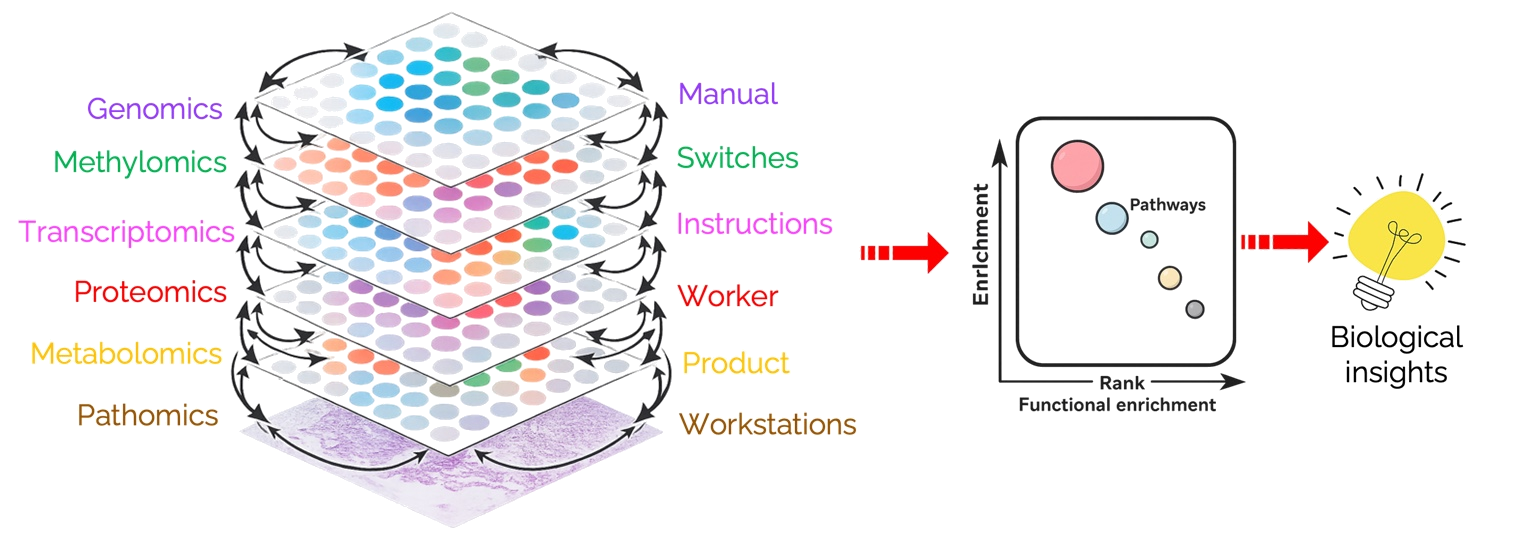
One way to think about multi-omics is as a layered biological system. Genomics represents the inherited biological instructions. Methylomics and other epigenomic marks act as regulatory switches that influence which instructions are accessible. Transcriptomics captures which genes are being actively read. Proteomics measures the proteins that carry out many cellular functions. Metabolomics reflects biochemical products and energetic state. Tissue-level image analysis adds spatial and structural context by measuring architecture, cell organization, vascular patterning, and regional heterogeneity.
In our research, multi-omics integration helps move from lists of altered molecules toward biological mechanisms. We use it to ask how maternal exposures reshape placental regulation across molecular, cellular, metabolic, vascular, and structural scales.
Horizontal integration
Horizontal integration combines datasets from the same biological layer across different samples, cohorts, time points, species, or experimental conditions. The goal is to identify patterns that are reproducible across contexts.
transcriptomics
transcriptomics
transcriptomics
For example, we can compare placental transcriptomic signatures from mouse models of maternal exposure with human placental datasets to identify shared inflammatory, metabolic, mitochondrial, or vascular programs. Similarly, image-derived tissue features from different cohorts can be integrated horizontally to test whether structural phenotypes are robust across datasets.
Vertical integration
Vertical integration combines different biological layers measured from the same samples or matched experimental groups. The goal is to connect regulation across scales, from molecular signals to tissue organization and phenotype.
For example, a maternal exposure may alter genes involved in nutrient transport, reduce the abundance of transport-related proteins, shift metabolite availability, and produce measurable changes in villous vascularization. Vertical integration connects these signals into one mechanistic model of placental adaptation or dysfunction.
Why both types of integration matter
Horizontal and vertical integration answer different questions. Horizontal integration asks whether a biological signature is consistent across datasets or contexts. Vertical integration asks how different molecular and tissue layers interact within the same biological system. Together, they help distinguish robust placental programs from dataset-specific effects and connect molecular regulation to structural and functional outcomes.
In our laboratory, this approach allows us to link maternal exposures with placental biology at multiple scales. We first analyze each data layer separately to identify exposure-associated changes. We then use pathway analysis, correlation networks, latent-factor models, multivariate integration, and machine-learning approaches to identify molecular and structural features that move together. This helps us prioritize pathways that are not only statistically altered, but also biologically connected across layers.
The final goal is to build interpretable models of placental function. Instead of treating transcriptomics, proteomics, metabolomics, and histology as separate datasets, we use them as complementary views of the same biological process. This allows us to identify candidate mechanisms, generate testable hypotheses, and better understand how the placenta adapts, compensates, or fails in response to maternal exposures.
- Hasin Y, Seldin M, Lusis A. Multi-omics approaches to disease. Genome Biology. 2017;18:83.
- Subramanian I, Verma S, Kumar S, Jere A, Anamika K. Multi-omics Data Integration, Interpretation, and Its Application. Bioinformatics and Biology Insights. 2020;14:1177932219899051.
- Rohart F, Gautier B, Singh A, Lê Cao KA. mixOmics: An R package for ‘omics feature selection and multiple data integration. PLOS Computational Biology. 2017;13(11):e1005752.
- Singh A, Shannon CP, Gautier B, et al. DIABLO: an integrative approach for identifying key molecular drivers from multi-omics assays. Bioinformatics. 2019;35(17):3055–3062.
- Argelaguet R, Arnol D, Bredikhin D, et al. MOFA+: a statistical framework for comprehensive integration of multi-modal single-cell data. Genome Biology. 2020;21:111.
- Bülow RD, Jäger PF, Isensee F, et al. Extending the landscape of omics technologies by pathomics. 2023.